Day 1 :
Keynote Forum
Hildegund C.J. Ertl
The Wistar Institute, USA
Keynote: Improving the outcome of T-cell immunotherapy of cancers by pre-conditioning T cell metabolism towards fatty acid catabolism
Time : 09:30-10:15

Biography:
Abstract:
Recent years have shown tremendous progress in active immunotherapy of cancers through tumor antigen (TA)-specific vaccines, adoptive T cell transfer or check point inhibitors. Nevertheless complete cures remain rare. TA-specific CD8+ T cells rapidly become exhausted once they enter solid tumors. This exhaustion, causing loss of tumor infiltrating CD8+ T cells during tumor progression, is in part cause by metabolic constrains, such as hypoxia and hypoglycemia, within the tumor microenvironment (TME). Lack of glucose forces T cells to switch from glycolysis for energy and biomass production to fatty acid catabolism. Most T cells are apparently unable to adjust their metabolism within the TME and as a consequence loose functions and eventually die. Pre-conditioning T cells during their initial activation in lymphatic tissues or their expansion in vitro towards fatty acid beta oxidation (FAO) preserves their functions and allows them to more effectively reduce tumor burden. CD8+ T cell metabolism can be modified by different types of manipulations such as knockdown of HIF-1a or other key factors of the glycolysis pathway to reduce the cells’ reliance on glucose. FAO can be enhanced directly by strengthening signaling through PPAR-a, the master regulator of lipid metabolism.
Keynote Forum
Jan O. Gordeladze
University of Oslo, Norway
Keynote: Impact of vitamin K2 on the prevention and treatment of various metabolic end endocrine diseases,discussing the implications of vitamin K2 in hormonal (G-protein) signaling, as well as integration with the effects of vitamins A and D3
Time : 10:15-11:00

Biography:
Dr. Jan O. Gordeladze, PhD (born 25th of April, 1950), holds a triple professor competence (medical biochemistry, physiology, and pharmacology), and is presently working as a professor at the Department of Biochemistry, Institute of Basic Medical Science, University of Oslo, Norway. He has previously been employed as the medical director of MSD, Norway, serving two years as a Fulbright scholar at the NIH, Bethesda, Maryland, USA, and from 2006-2009 also being employed as associate professor at the University of Montpellier, France. He is a member of the Norwegian Stem Cell Center, and his research has over the past 7-10 years been devoted to differentiation of osteochondral cells from stem cells focusing on the impact of transcription factors and microRNA species constituting regulatory loop interactions with functional target genes. He has published more than 120 scientific articles, reviews/book chapters and presented more than 250 abstracts/posters/talks at conferences world wide. Dr. Gordeladze has served as a Fulbright Scholar at the National Institute of Health, Bethesda, Washington DC during the years 1990-91.
Abstract:
To be updated
Keynote Forum
Joel I. Osorio
Westhill University, Mexico
Keynote: RegenerAge System: Therapeutic effects of combinatorial biologics(mRNA and Allogenic MSCs) with a spinal cord stimulation system on a patient with spinal cord section towards fatty acid catabolism
Time : 14:00-14:40

Biography:
Abstract:
- Molecular Medicine | Molecular Biology | Molecular pathology | Biochemistry | Biomarkers & Diagnostics Molecular Drug Designing | Molecular Diagnostics | Molecular Modeling and Dynamics | Molecular Virology
Session Introduction
Xiong Wen
Sichuan Provincial People’s Hospital, China
Title: Hormonal manipulation induces differentiation of pancreatic progenitor cells into insulin secreting islet β-cells
Time : 14:40-15:05

Biography:
I has been engaged in ultrasonic diagnosis for 12 years, mastered the abdomen and superficial tissue disease is a common and rare disease, specialize in fetal abnormalities of prenatal ultrasound diagnosis and the ultrasonic diagnosis of children abdominal and superficial tissue diseases.
Abstract:
Diabetes is a kind of metabolic disease, which causes considerable morbidity in the world. Although pancreas transplantation and islet transplantation has prominent future, we still confront the main difficulty of organ shortage. Thus, a primary and fundamental effective therapy for diabetes is to develop ways to increase beta cell numbers.Here, we reported that under the stimuli of homones, pancreatic duct epithelial cells, also known as pancreatic progenitor cells, could be differented into insulin-secreting islet β-cells.
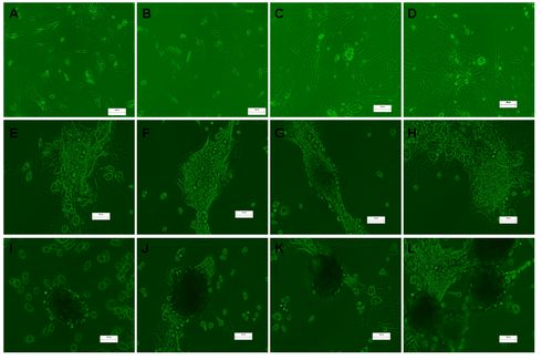
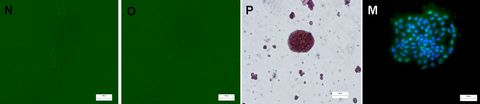
In this study, we collected pregnant rat serum and added to the culturing medium of isolated rat pancreatic duct epithelial cells. After 7 days of culturing, the pancreatic progenitor cells will be aggregrated (Figure 1). Then we compared the gender difference of the pancreatic progenitor cells, and also the dosage of pregnant serum on the efficiency of differentiation. As observed in Figure 2, all cells treated with pregnant serum experienced expansion (A-D), aggregation (E-H), and islet-like cells formation (I-L) stages. Higher concentration of pregnant serum treated cells (L,J) generate more islet-like cells compared to those of lower ones (I, K). Pancreatic duct epithelial cells isolated from female rats formed larger islet-like sphere compared to those of male ones. However, pancreatic duct epithelial cells cultured with FBS could not form islet-like cells (N, low concentration of FBS control; O, high concentration of FBS control). Then the differented islet-like cells were determined by dithizone staining (P) and aggregated pancreatic duct cells were determined by insulin staining (M). Judged from these two pictures, no matter the aggregrated cells or the sphere-shaped ones are capable of secreting insulin.
In conclusion, the pancreatic progenitor cells could be differented to insulin-secreting islet β-cells by the pregnant serum, which indicats the therapeutic potential of hormone therapy in preventing and/or treating diabetes.
Figure 1:
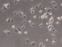
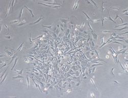
Figure 2:
Ying Liu
Capital Medical University,China.
Title: Effect of local endometrial injury in proliferative vs. luteal phase on IVF outcomes in unselected subfertile women undergoing in vitro fertilization
Time : 15:05-15:30

Biography:
Ying Liu has completed her postdoctoral studies from Yale University School of Medicine. She is a professor and PhD supervisor. She has published more than 50 papers in journals.
Abstract:
Mechanical endometrial injury prior to IVF has been suggested as a means to increase implantation rates by improving endometrial receptivity. However, the effects of endometrial injury in proliferative vs. luteal phase have not been studied before. This study aimed to explore whether endometrial injury in the proliferative phase of the preceding cycle before in vitro fertilization/embryo transfer (IVF-ET) improves the clinical outcomes in unselected subfertile women compared with injury in luteal phase. In our study, a group of 142 patients were randomized into four groups: injury group (group A: endometrial injury in proliferative phase, n=38; group B: endometrium injury in luteal phase, n=32), and non-injury group as control (group C: non-injury in proliferative phase, n=36; group D: non-injury in luteal phase, n=36). Patients in injury groups underwent endometrial injury in either proliferative phase or luteal phase in the preceding cycle before IVF treatment. Clinical outcomes including implantation, pregnancy, and live birth rates were analyzed among the four groups. As result, the baseline characteristics of the four groups including age, body mass index, duration, type and causes of infertility were similar. There were no significant differences in implantation, clinical pregnancy or live birth rates between injury group and non-injury group. Moreover, there were also no significant differences in implantation, clinical pregnancy, or live birth rates in injury in proliferative phase compared with luteal phase. In conclusions, endometrial injury in the cycle preceding IVF of unselected subfertile women does not increase implantation, clinical pregnancy, or live birth rates. Furthermore, there is no significant difference in clinical outcomes between endometrial injury in the proliferative phase and injury in the luteal phase.
Radwa Hussein Mohamed Ghoraba
Alexandria University, Egypt
Title: Quantitative analysis of human herpes virus 6 dna in patients treated for acute leukemia
Time : 15:30-15:55

Biography:
Radwa H Ghoraba a Pharmacist, graduated from Faculty of Pharmacy and Drug Manufacturing, Pharos University 2012, Alexandria, Egypt. She has a Master’s degree in Diagnostic and Molecular Microbiology, Medical Research Institute, Alexandria University 2017, Egypt. Her involvement in research has given her first-hand exposure to the process of active scientific research, resulted in incredible research experiences, and instilled in her a passion for science and exploration. She is interested in improving public health through research. She is really interested in the theme of the 3rd International Conference on Molecular Medicine and Diagnostics which is; Exploring the Modern Innovations in the Field of Molecular Medicine and Diagnostics.
Abstract:
Viral infections are important causes of morbidity and mortality for patients with a hematological malignancy, but the true incidence and consequences of viral infections for these patients who undergo conventional non transplant therapy are inadequately defined. Viral infections in hematological patients may result from reactivation of latent infection or, rarely, from acquisition of a new infection. Thus, screening of patients with hematological malignancies for HHV-6 might be considered mandatory. The aim of this study was to evaluate a possible association between Human Herpesvirus-6 (HHV-6) infection and acute leukemia in adults after receiving chemotherapy treatment for acute leukemia. The patients were divided into two main groups according to the type of leukemia, All patients with newly diagnosed acute leukemia were subjected to history taking, complete clinical examination and routine laboratory investigations. Peripheral blood samples (Whole blood specimens) were collected from all patients for quantitative determination of HHV6 DNA viral load by Taqman probe technique (real time PCR) at day 0 and day 100 of induction chemotherapy after being extracted on day of sampling. Data were fed to the computer and analyzed using IBM SPSS software package version 20.0. (Armonk, NY: IBM Corp). The results argued against an etiological relationship between HHV-6 infection and the genesis of acute leukemia in adults, however, it supports the hypothesis of viral latency and the possibility of virus reactivation in immunocompromised hosts. The possible presence of HHV-6 as an associated or a putative causative agent in leukemia should however be considered.
Doaa Ali Abdelmonsif
Alexandria University Alexandria, Egypt
Title: Targeting AMPK, mTOR and βCatenin by Combined Metformin and Aspirin Therapy in HCC: AnAppraisal to Egyptian HCC Patients
Time : 16:10-16:35

Biography:
Doaa A. Abdelmonsif has completed her MD in Medical Biochemistry since 2011 from Alexandria Faculty of Medicine, Egypt. She is a co-director of the molecular biology laboratory of the Center Of Excellence for Research In Regenerative Medicine and It is Applications, Alexandria Faculty of Medicine, Egypt. She has several publications in the field of molecular medicine and targeted drug delivery for treatment of cancer and neurodegenerative diseases. She serves as a reviewer for several reputed journals.
Abstract:
Hepatocellular carcinoma (HCC) is an expanding health problem with a great impact on morbidity and mortality both in Egypt and worldwide. Recently, metformin and aspirin showed a potential anticancer effect on HCC although their mechanism(s) isn't fully elucidated. To investigate the anti-proliferative effects of combined metformin/ aspirin treatment against HCC, HepG2 cells were exposed to increasing concentrations of metformin, aspirin and combined treatment, and MTT assay was performed. Caspase-3 activity, cell cycle analysis, protein expression of phosphorylated AMP-activated protein kinase (pAMPK) and mammalian target of rapamycin (mTOR) proteins were assessed. Furthermore, the expression and localization of β-catenin protein was assessed by immunocytochemistry (ICC). Finally, protein expression of pAMPK, mTOR and β-catenin was assayed in Egyptian HCC and cirrhotic tissue specimens. Results showed that metformin/aspirin combined treatment had a synergistic effect on cell cycle arrest and apoptosis induction via down-regulation of AMPK activation and mTOR protein expression. Additionally, metformin/aspirin combined treatment enhanced cell-cell membrane localization of β-catenin expression in HepG2 cells, which might inhibit metastatic potential of HepG2 cells. In Egyptian HCC specimens, pAMPK, mTOR and β-catenin proteins showed a significant expression compared to cirrhotic controls. In conclusion, combined metformin/aspirin treatment could be a promising therapeutic strategy for HCC and specifically Egyptian HCC patients.
Abdulkarim Jamal
Warwick university- Nuneaton, United Kingdom
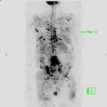
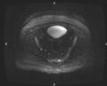
Title: Whole body diffusion weighted MRI in the detection of skeletal metastasis. our experience with 1.5 and 3 Tesla MRI
Time : 16:35-17:00

Biography:
Abstract: