Abdulkarim Jamal
Warwick university- Nuneaton, United Kingdom
Title: Whole body diffusion weighted MRI in the detection of skeletal metastasis. our experience with 1.5 and 3 Tesla MRI
Biography
Biography: Abdulkarim Jamal
Abstract
Introduction:
Metastatic bone disease is a common manifestation of advanced cancers particularly breast, prostate and lung. Currently many hospitals rely on traditional imaging technique such as bone scan, CT scan and more recently PET CT scan for detection of metastases and assessment of tre
Whole-body Diffusion Weighted Imaging (WB-DWI) is emerging as a promising bone marrow assessment tool for detection and therapy monitoring of bone metastases.
Advantages of WB-DWI include the fact that no ionizing radiation is administered and no injection of isotopes or any contrast medium is necessary.
Importantly, whole-body skeletal examinations are possible in reasonably short data acquisition times.
Methods:
110 whole body MRI examinations using 1.5 and 3 Tesla MRI and the image findings in skeletal metastases and comparison with other imaging modalities.
Results:
WB-MRI outperforms bone scans + targeted x-rays in detecting bone metastases and performs as well as CT for lymph node evaluation in prostate cancer.
FDG-PET and WB-DWI; both are more accurate than CT and bone scans for detecting bone metastases.
WB-DWI used alone has equal performance to FDG-PET for detecting primary tumours &soft tissue metastases
DW-MRI needs to be combined with morphologic sequences to improve specificity.
Conclusions:
Whole body MRI is promising new technique in the detection and assessment of treatment response in skeletal metastases in many malignancies. It has very high sensitivity and specificity, its relatively quick to perform and does not need the injection of contrast agent or the use of ionising radiation.
Padhani AR, et al. Therapy monitoring of skeletal metastases with whole body diffusion MRI. J Magn Rson Imaging. 2014;39(5):1049-78
Yang HL, et al. Diagnosis of bone metastases: a meta-analysis comparing 18FDG PET, CT, MRI and bone scintigraphy Eur Radiol. 2011; 21:2604-17
Figure1: Coronal reformat MIP inverted b900 image depicting wide spread skeletal metastases in a patient with prostate cancer
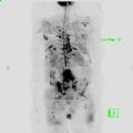
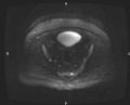
Figure 2: Axial b900 image of the pelvis depicting 2 small lesions(bright) with restricted diffusion in keeping with Myeloma deposits ( not visible on plain film)